


Confocal vs Widefield Microscopy: Which Imaging Method Fits Your Research Workflow?
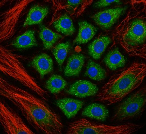
Choosing between confocal and widefield microscopy can affect image contrast, acquisition speed, specimen exposure and the quality of the resulting data. Both techniques are widely used in life science research, but they are suited to different imaging conditions.
For laboratories assessing advanced microscopy technologies, the right choice depends on specimen thickness, fluorescence distribution, required imaging depth and the scientific question being investigated.
How Does Widefield Microscopy Work?
Widefield microscopy illuminates a broad field of the specimen and captures emitted or transmitted light across the image simultaneously.
It can be effective for thin specimens, routine fluorescence imaging and applications where rapid image acquisition is important.
Potential advantages include:
-
Rapid image acquisition across a wide field
-
Relatively straightforward imaging workflows
-
Suitability for many thin specimens
-
Efficient observation of suitable fluorescent samples
Important limitation: In thicker specimens, out-of-focus fluorescence may contribute to background haze and reduce contrast.
How Does Confocal Microscopy Work?
Confocal microscopy typically uses focused laser illumination and a spatial pinhole to reject much of the out-of-focus light reaching the detector.
This optical sectioning capability can improve contrast and enable images to be acquired from selected depths within a specimen.
Potential advantages include:
-
Improved contrast in many fluorescent specimens
-
Optical sectioning for three-dimensional imaging
-
Better separation of structures at different depths
-
Support for selected thick-sample and cellular imaging applications
Important limitation: Acquisition may be slower than widefield imaging, depending on the system and settings. Laser exposure can also cause photobleaching or phototoxicity, particularly in sensitive live specimens.
Confocal vs Widefield Microscopy: Key Differences
| Factor | Widefield microscopy | Confocal microscopy |
|---|---|---|
| Illumination | Broad field illumination | Typically focused laser illumination |
| Out-of-focus light | Can contribute significant background | Much of it is rejected optically |
| Thin specimens | Often well suited | Can be suitable |
| Thick specimens | Background may limit contrast | Optical sectioning can improve contrast |
| Acquisition | Often fast for full-field capture | Depends on scan speed and image settings |
| Equipment complexity | Often simpler | Typically more specialised |
| Best selection factor | Sample and workflow suitability | Need for optical sectioning and contrast |
Neither method is universally superior. The specimen and research objective should determine the choice.
Which Technique Is Better for Fluorescence Imaging?
For thin fluorescent specimens, widefield microscopy may provide adequate contrast with efficient acquisition.
For specimens containing structures at different depths, confocal microscopy can help distinguish features by reducing out-of-focus background. However, its advantage depends on the specimen, labelling and optical configuration.
For low-light experiments, evaluate the total system, including illumination, detector sensitivity, exposure time and potential photodamage.
When Should You Consider Confocal Microscopy?
Confocal microscopy may be worth evaluating when your research requires:
-
Optical sections through a specimen
-
Improved contrast in samples with out-of-focus fluorescence
-
Three-dimensional reconstruction from image stacks
-
Observation of labelled structures at different depths
-
More selective fluorescence imaging
If the main requirement is to capture images of thin, well-prepared samples quickly, widefield microscopy may remain a practical choice.
What About Super-Resolution Microscopy?
Confocal microscopy improves optical sectioning but does not, by itself, overcome the conventional diffraction limit.
When the research requires resolving selected structures at finer spatial scales, techniques such as STED microscopy or other super-resolution approaches may be relevant.
Explore Super-Resolution Microscopy to investigate this separate class of imaging technology.
How to Select the Right Microscopy System
Before comparing systems, document the specimen thickness, fluorophores, required resolution, imaging depth, acquisition speed and intended analysis.
Also consider whether the system will require a compatible scientific camera, image-acquisition software or additional accessories.
Explore Microscopy Applications at DSS Imagetech to review the broader range of microscopy techniques.
Frequently Asked Questions
Is confocal microscopy always better than widefield microscopy?
No. Confocal microscopy can improve optical sectioning and contrast in suitable specimens, while widefield microscopy can be faster and effective for thin samples.
Can widefield microscopy be used for three-dimensional imaging?
Image stacks can be acquired by changing focus, but out-of-focus light may reduce contrast in thicker specimens. The suitability depends on the sample and imaging method.
Does confocal microscopy improve resolution?
Confocal microscopy can improve contrast and optical sectioning. Its resolution benefits depend on the imaging conditions, and it does not automatically provide super-resolution beyond the conventional diffraction limit.
How do I choose between confocal and widefield systems?
Consider specimen thickness, background fluorescence, imaging depth, required speed, phototoxicity and the level of structural detail needed.

 English
 French
 Spanish
 Portuguese
 Deutsch
 Turkish
 Dutch
 Italiano
 Russian
 Romaian
 Portuguese (Brazil)
 Greek